



 English
 Español


Most people carry a pair of breasts through their entire adult lives without ever thinking much about what is actually inside them. Yet the breast is one of the more architecturally sophisticated structures in the human body — a layered system of glands, ducts, fat, connective tissue, nerves, and blood vessels that changes across a lifetime in response to hormones, age, pregnancy, and lactation.
Understanding that architecture is not just academically interesting; it gives context to mammography reports, clinical examinations, and the wide natural variation in breast size, shape, and density that research consistently documents.
This guide works through the breast from the outside in — skin and surface landmarks first, then the internal tissue compartments, the glandular machinery, the structural supports, and finally the nerve and blood supply that keeps everything functioning.
"The breast is not a single tissue but a composite organ — fatty, glandular, and fibrous compartments each with distinct roles that shift dramatically across a woman's reproductive life."
— Gray's Anatomy for Students, 4th ed., Drake et al., 2019
Surface Anatomy: What You Can See and Feel
The breast sits on the anterior chest wall, overlying the pectoralis major muscle and, to a lesser extent, the serratus anterior. In most women, breast tissue extends from roughly the second rib to the sixth or seventh rib vertically, and from the lateral edge of the sternum to the mid-axillary line horizontally. An extension of tissue, called the axillary tail of Spence, reaches toward the armpit and is clinically significant because it contains glandular tissue that can be affected by the same conditions as the main breast mass (National Library of Medicine, Anatomy of the Breast).
The skin covering the breast is ordinary stratified squamous epithelium, but two surface features deserve specific attention: the areola and the nipple.
The areola is the pigmented circular zone surrounding the nipple. Its diameter varies considerably among individuals — studies using standardised photographic measurement in adult women have recorded average diameters in the range of 3.5 cm to 4.5 cm (roughly 1.4 to 1.8 in), with wide variation considered within the studied range (Sanchez et al., Aesthetic Plastic Surgery, 2017). The areola contains Montgomery glands, small sebaceous structures that appear as raised bumps — typically 10 to 15 per areola — which secrete an oily fluid thought to lubricate the nipple during breastfeeding and produce subtle scent cues for newborns (Doucet et al., Chemical Senses, 2009).
The nipple (medical term: papilla mammae) is a cylindrical projection at the center of the areola. It is perforated by 15 to 25 lactiferous duct openings through which milk exits the breast during lactation. Nipple position, projection, and orientation vary widely. Inverted nipples — where the nipple retracts inward — occur in an estimated 10–20% of women and most are a variation of typical development rather than a clinical problem (Alexander et al., British Journal of General Practice, 1999).
Did You Know?
Montgomery glands — those small bumps on the areola — are a hybrid between a sebaceous gland and a mammary gland. Research published in Chemical Senses found that newborns orient preferentially toward secretions from these glands, suggesting they play a role in guiding feeding behaviour from the very first hours of life.
Inside the Breast: Three Tissue Compartments
Cut through a breast surgically and you encounter three principal tissue types arranged in a way that varies considerably from person to person and changes across a lifetime.
1. Glandular Tissue (Parenchyma)
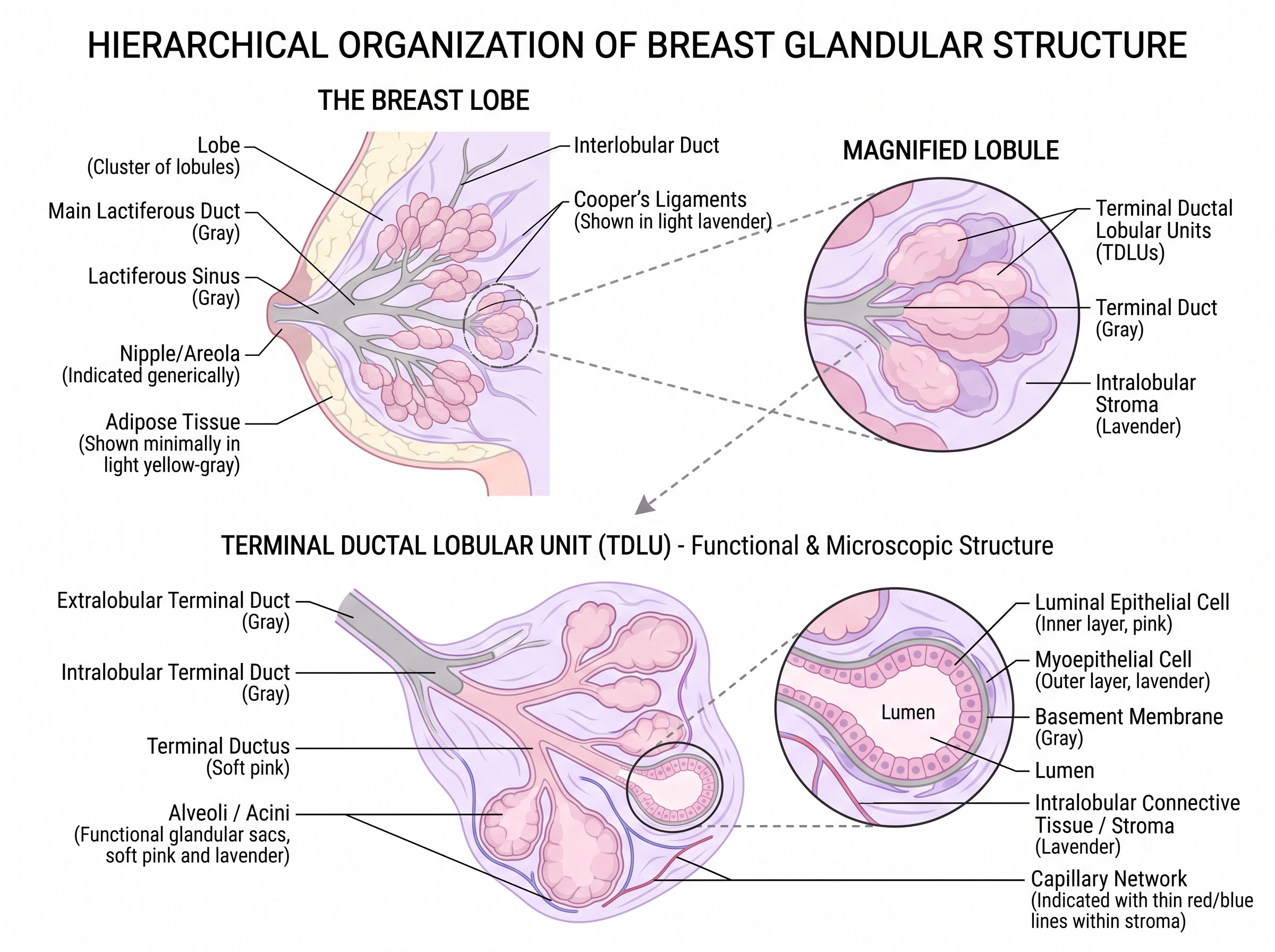
The functional core of the breast is its glandular apparatus — the machinery that can produce milk. This system is organised hierarchically. At the top level, the breast contains 15 to 20 lobes, each a self-contained gland arranged roughly in a wheel-spoke pattern around the nipple. Each lobe drains into its own lactiferous duct, which widens just behind the areola into a small reservoir historically called the lactiferous sinus (though modern anatomy texts note this dilation is less pronounced than older descriptions suggested; Going & Moffat, Journal of Anatomy, 2004).
Each lobe is further divided into lobules, and each lobule contains clusters of rounded secretory units called terminal ductal lobular units (TDLUs). The TDLU is the functional building block of the breast — it is where milk is synthesised during lactation, and it is also the site where most breast carcinomas originate (Russo & Russo, Microscopy Research and Technique, 2004).
Outside of pregnancy and lactation, the glandular component is relatively compact and non-secretory. Hormonal shifts during the menstrual cycle, however, cause cyclic changes in ductal and lobular tissue — which is why breast tenderness commonly tracks with the luteal phase.
2. Adipose Tissue (Fat)
Surrounding and interpenetrating the glandular tissue is adipose — fatty — tissue, and in most adult women, this is the largest single component of breast volume. The ratio of fat to glandular tissue changes substantially with age: younger women tend to have proportionally more glandular tissue (termed dense breasts on mammography), while after menopause the glandular component involutes and adipose tissue predominates (Boyd et al., New England Journal of Medicine, 2007).
This ratio matters clinically because dense breast tissue appears white on a mammogram — the same shade as tumours — making detection more challenging. The National Cancer Institute's resource on breast density offers accessible detail on how density classifications are used in screening contexts.
3. Stromal / Connective Tissue
Woven throughout both the glandular and fatty compartments is a fibrous stromal framework — collagen, elastin, and a ground substance of proteoglycans. This stroma is not merely scaffolding; it is an active biological environment that communicates with epithelial cells via signalling molecules and plays a documented role in breast cancer biology (Bhowmick et al., Nature, 2004).
Cooper's Ligaments: The Internal Support System
The breast has no internal muscle of its own and contains no cartilage or bone. Its structural integrity depends largely on Cooper's ligaments (technically, suspensory ligaments of the breast), named after the British surgeon Sir Astley Cooper who described them in 1840. These are fibrous bands that run from the dermis of the overlying skin down through the breast tissue to attach to the deep fascia overlying the pectoral muscle, effectively suspending the breast and maintaining its contour.
Cooper's ligaments are not static. They gradually stretch and thin with age, gravitational load, and repeated cycles of expansion and contraction (as during pregnancy and weight changes), which contributes to the ptosis — drooping — many women observe over decades. Research using ultrasound has confirmed their structural role and their progressive laxity with age (Hussain et al., British Journal of Radiology, 2016).
Clinically, invasion of Cooper's ligaments by a tumour causes them to shorten, which produces a characteristic skin dimpling or puckering on the breast surface — a physical sign taught in medical schools worldwide as a potential indicator warranting investigation.
Clinical Note
Skin dimpling on the breast — particularly when it appears in a new or unexplained way — is a physical sign that warrants prompt medical evaluation. It can arise when a tumour invades and shortens Cooper's ligaments, pulling the skin inward. This article is educational only; always consult a qualified healthcare provider about any changes to breast tissue.
Blood Supply: Where the Breast Gets Its Nutrients
The breast receives blood from multiple arterial sources, a redundancy that reflects its metabolic importance — particularly during lactation, when blood flow to the breast increases dramatically.
The primary contributors are:
- Internal thoracic artery (internal mammary artery): Perforating branches pass through the intercostal spaces — particularly the second, third, and fourth — and supply the medial and central breast. This is typically the dominant supply, estimated to account for roughly 60% of breast perfusion (Hamdi et al., Annals of Plastic Surgery, 2009).
- Lateral thoracic artery: A branch of the axillary artery supplying the upper outer quadrant — the quadrant that, incidentally, contains the greatest density of glandular tissue and where the majority of breast carcinomas are detected.
- Thoracoacromial artery (pectoral branch): Contributes to the upper breast.
- Intercostal arteries (lateral branches): Contribute to the lateral and inferior breast.
Venous drainage roughly mirrors the arterial supply, draining toward the axillary vein and internal thoracic vein. This venous anatomy is relevant to understanding how breast cancer cells can enter systemic circulation.
Table 1 — Principal Blood Supply to the Breast
Lymphatic Drainage: Why Surgeons Pay Close Attention
The breast has a rich lymphatic network, and its drainage pathways are studied intensively in the context of breast cancer spread. The majority of lymph from the breast — approximately 75% — drains to the axillary lymph nodes, a group of 20 to 40 nodes located in the armpit. The remainder drains to the internal thoracic (parasternal) nodes along the sternum, and a small proportion to posterior intercostal nodes (Suami et al., Plastic and Reconstructive Surgery, 2008).
The sentinel lymph node — the first node a tumour cell would encounter on its journey out of the breast — is identified and sampled during breast cancer surgery precisely because of this predictable drainage hierarchy. If the sentinel node is clear of cancer cells, surgeons can often avoid removing the full axillary chain, significantly reducing the risk of lymphoedema.
Nerve Supply: Sensation and Its Variation
Breast sensation is carried primarily by branches of the intercostal nerves (T2 through T6), with the lateral and anterior cutaneous branches supplying the main skin envelope. The nipple and areola receive their most important sensory supply from the lateral cutaneous branch of the fourth intercostal nerve (T4), which approaches the nipple from the lateral side — a fact that surgeons performing breast reduction or augmentation must account for to preserve nipple sensation (Sarhadi et al., Journal of the Royal College of Surgeons of Edinburgh, 1996).
Sensation in the breast and nipple varies considerably between individuals. Research using quantitative sensory testing has found substantial differences in touch thresholds and vibration sensitivity across the breast surface, with the nipple-areola complex generally being the most sensitive region (Schlenz et al., Plastic and Reconstructive Surgery, 2000). Many women report that nipple sensitivity changes across the menstrual cycle, and that pregnancy and lactation can produce marked changes — sometimes increased sensitivity, sometimes temporary reduction — that typically normalise after weaning.
Nipple erection (the technical term is nipple erection reflex) is mediated by sympathetic nerve fibres acting on smooth muscle within the nipple. It is triggered by tactile stimulation, cold, and sexual arousal — a reflex shared, incidentally, with male nipples, which are anatomically homologous structures.
Cultural Insight
Breasts Through History
Ancient Greek anatomists grouped the breast with reproductive organs, believing it was directly connected to the uterus via blood vessels — a theory that persisted well into the Renaissance.
Sir Astley Cooper's 1840 work On the Anatomy of the Breast — illustrated with extraordinary precision — remained the definitive anatomical reference for over a century. Many of his observations about the ligaments that now bear his name remain accurate today.
In the 20th century, the development of mammography transformed breast anatomy from a surgical curiosity into a radiological map — making internal tissue density a clinically measurable and medically significant variable.
Documented Variation: Size, Shape, and Density
Population studies consistently show that breast morphology — size, shape, volume, and tissue composition — varies widely and that most of this variation falls within what researchers consider the studied range of human anatomy. A large-scale 3D surface imaging study of over 400 women by Losken et al. documented substantial variation in breast volume, projection, and ptosis grade even within age-matched cohorts (Plastic and Reconstructive Surgery, 2005).
Breast volume measurements vary between roughly 100 mL to over 1,500 mL (0.4 to 6.3 cups) per breast in adult women, with the majority falling between 400 and 800 mL (Katch et al., Annals of Human Biology, 1980). Asymmetry between left and right breasts is the rule rather than the exception — studies suggest over 88% of women have some measurable volumetric asymmetry, with the left breast being larger in most cases (Scutt et al., The Breast Journal, 2006).
Understanding how size and breast composition relate is also relevant reading alongside our article on Breast Size Measurement: Methods, Standards and What the Data Shows. For readers interested in how hormones drive these structural changes across the lifespan, see our overview on Hormones and Breast Development: Puberty to Menopause.
In Brief
- The breast contains three tissue compartments: glandular (lobes and ducts), adipose (fat), and fibrous stromal tissue.
- The functional unit is the terminal ductal lobular unit (TDLU) — where milk is made and where most carcinomas originate.
- Cooper's ligaments suspend the breast from the skin to the deep fascia; their gradual laxity contributes to ptosis over time.
- The primary blood supply comes from the internal thoracic artery (~60%) and lateral thoracic artery.
- About 75% of lymph drains to axillary nodes — the basis of sentinel node biopsy in breast cancer surgery.
- Nipple sensation is primarily carried by the lateral cutaneous branch of the T4 intercostal nerve.
- Breast asymmetry is present in over 88% of women; wide variation in size, density, and shape is within the studied range.
By the Numbers
Breast Anatomy: Common Questions Answered
Breast anatomy is not a single fixed picture but a living architecture that remodels across decades in response to hormones, reproduction, ageing, and individual genetics. Knowing what the different layers are, how they are supported, and what systems supply and drain them makes the language of mammography reports, surgical consultations, and clinical breast examinations considerably more legible. Research in this area continues to refine our understanding — particularly around the biological role of the stroma and the mechanics of lymphatic spread — and keeping up with that science remains one of the clearest arguments for broad anatomical literacy.
Disclaimer
- All content on this website—including articles, educational materials, and interactive calculators—is for informational, educational, and entertainment purposes only. The calculations, percentiles, and outputs generated by tools on this site are based on general statistical data and mathematical models; they do not constitute medical data, a clinical assessment, or a diagnosis.
- Nothing contained on this website is a substitute for professional medical advice, diagnosis, or treatment. Always seek the guidance of a qualified healthcare professional or urologist with any questions you have regarding physical development, anatomy, or health conditions. Reliance on any information or tools provided by this website is solely at your own risk.
