



 English
 Español


Six weeks after fertilization, a human embryo is roughly 1.2 centimetres (0.5 inches) from crown to rump, and there is nothing about its lower body that would let anyone tell one from another. There is a ridge of tissue that could become a testis or an ovary. There is a small bud. There are two swellings on either side of it. And, unusually for a body plan that will eventually pick one option, there are two complete sets of internal plumbing, laid down side by side, both fully intact.
What happens next is one of the more orderly sequences in human biology. Over the following eight weeks, a short list of hormonal signals arrives in a fixed order, each doing one specific job, and the shared starting material is either reworked or left alone. The companion piece to this article, The Shared Blueprint: How One Fetal Template Becomes Male or Female, covers what that starting material is. This article traces the signals — what arrives, when, and what each one is responsible for.
In Brief
- Every human embryo starts with the same gonad, the same small bud of tissue, and both sets of internal ducts.
- Three signals do most of the work: anti-Müllerian hormone, testosterone, and dihydrotestosterone (DHT). Each has a separate job.
- The female pathway is not a fallback. Research has identified genes that actively build and maintain the ovary.
- Timing matters as much as quantity. Several of these signals only count during narrow windows measured in weeks.
Weeks 4 to 7: Everything Is Still Optional
The genital ridges appear on the back wall of the embryo at around four to five weeks after fertilization. Developmental biologists call the resulting organ the bipotential gonad, and the name is accurate: at this stage the tissue carries the cellular machinery for both outcomes, and published work on gonadal development describes it as genuinely undecided rather than merely unlabelled (Wilhelm, Palmer & Koopman, Physiological Reviews, 2007).
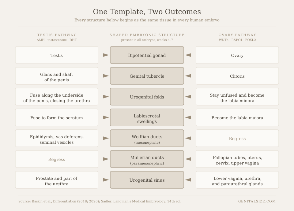
Two paired ducts run alongside it. The Wolffian ducts, if kept, can become the epididymis, the vas deferens and the seminal vesicles. The Müllerian ducts, if kept, can become the fallopian tubes, the uterus, the cervix and the upper vagina. Every embryo at this stage has both. On the outside, three structures sit ready: the genital tubercle, a pair of urogenital folds, and a pair of labioscrotal swellings. Studies of human external genital development report that these are indistinguishable across embryos until around week nine (Baskin et al., Differentiation, 2018).
One point of precision, because published sources differ: the weeks quoted throughout this article are counted from fertilization. Pregnancy is more commonly dated from the last menstrual period, which runs about two weeks ahead. A structure described here as forming at week nine is at roughly week eleven on a standard pregnancy calendar.
The Switch, and the Six Days That Follow
The trigger is a single gene. In 1990 a research team identified SRY as the sex-determining region on the Y chromosome, publishing the finding in Nature. Work on human tissue has since placed the window of SRY expression in the gonad at roughly 41 to 44 days after fertilization — a burst lasting only a few days (Hanley et al., Mechanisms of Development, 2000).
SRY does not build anything itself. It raises the activity of a second gene, SOX9, and that pushes a population of supporting cells in the gonad to become Sertoli cells. Once Sertoli cells exist, the tissue has committed, and everything downstream follows from what those cells and their neighbours then secrete. This is the part that surprises people: the chromosome's direct contribution is brief and narrow. Almost all of the anatomy is built afterwards, by hormones, and by hormones that could in principle come from anywhere.
The chromosome's direct contribution lasts a few days. Almost every structure that follows is built by hormones — which is why the timing of those hormones turns out to matter so much.
Signal One: Anti-Müllerian Hormone, the Demolition Order
The first hormone out of the gate is not testosterone. From around week seven, the newly formed Sertoli cells begin producing anti-Müllerian hormone, usually shortened to AMH. Its function is unusually specific for a hormone: it causes the Müllerian ducts to break down, a process research describes as largely complete by around week ten (Josso et al., Molecular and Cellular Endocrinology, 2001).
AMH removes the option. It does not add a structure. If AMH is absent or the tissue cannot respond to it, the Müllerian ducts stay — regardless of what testosterone is doing elsewhere in the same embryo. The two signals run on separate tracks, which is why developmental biologists treat them as independent jobs rather than two aspects of one process.
Signal Two: Testosterone, the Preservation Order
At around week eight, a second cell population in the testis — the Leydig cells — begins producing testosterone. Measurements of hormone levels in fetal tissue put the peak between roughly weeks eleven and seventeen, after which output falls away for the rest of the pregnancy (Reyes et al., Journal of Clinical Endocrinology & Metabolism, 1974).
Testosterone's job at this stage is the mirror image of AMH's: it keeps the Wolffian ducts from breaking down and drives them to develop into the epididymis, the vas deferens and the seminal vesicles. It works locally, at short range, on the duct sitting next to the testis producing it. Testosterone alone, though, does very little to the structures on the outside. For those, the body performs a chemical conversion first.
Signal Three: DHT, and Why the Body Bothers Converting
An enzyme called 5-alpha-reductase type 2, present in the tissue of the developing external genitalia, converts testosterone into dihydrotestosterone. DHT binds the androgen receptor more tightly and releases it more slowly than testosterone does, which makes it a stronger signal at the same concentration (Grino, Griffin & Wilson, Endocrinology, 1990). The body is not making a different message. It is making a louder one, in the exact place it is needed.
From around week nine through week fourteen, DHT drives three changes at once. The genital tubercle lengthens rather than staying compact. The urogenital folds fuse along the midline, closing over to enclose the urethra along the underside of the penis. The labioscrotal swellings fuse to form the scrotum. DHT also drives the prostate to bud from the urogenital sinus. The classic account of this division of labour — testosterone for internal ducts, DHT for external structures and prostate — was set out by Wilson, George and Griffin in Science in 1981 and has held up since.
Growth continues after the structural work is done. The external anatomy is essentially built by week fourteen; the third trimester adds size, not new parts.
Table 1. The sequence, by week after fertilization
| Week | What happens | Signal responsible |
|---|---|---|
| 4–5 | Genital ridges form; the gonad is bipotential | — |
| 6–7 | Both duct systems present; external structures identical in all embryos | — |
| 6–7 | SRY switches on briefly; Sertoli cells form | SRY → SOX9 |
| 7–10 | Müllerian ducts break down | Anti-Müllerian hormone |
| 8–14 | Wolffian ducts preserved and developed | Testosterone |
| 9–14 | Tubercle lengthens; folds and swellings fuse; prostate buds | DHT |
| 8–12 | In the absence of AMH, Müllerian ducts fuse into uterus and upper vagina | WNT4 / RSPO1 pathway |
| ~20 | Egg cell count in the fetal ovary reaches its lifetime maximum | — |
Weeks are counted from fertilization, not from the last menstrual period. Ranges are approximate and vary between published sources.
The Female Sequence Is Not the Absence of the Male One
For most of the twentieth century, textbooks described female development as what happens when nothing happens — the "default" pathway. That description has not survived the genetics.
Researchers have identified a set of genes that actively build the ovary and keep it an ovary. WNT4 was shown in 1999 to be required for ovarian development in mammals (Vainio et al., Nature). RSPO1 was added to the list in 2006 (Parma et al., Nature Genetics). Most striking is FOXL2: a 2009 study in Cell reported that switching FOXL2 off in an adult mouse ovary caused the supporting cells to begin taking on the character of testis cells (Uhlenhaut et al.). Maintaining an ovary, in other words, is an ongoing job rather than a decision made once and forgotten.
The reproductive tract follows. Without AMH, the Müllerian ducts persist and their lower portions fuse along the midline, forming the uterus and cervix, with the upper vagina developing from the same material; the unfused upper sections remain as the fallopian tubes (Robboy et al., Differentiation, 2017). Without DHT, the external structures keep their original arrangement: the genital tubercle becomes the clitoris, the urogenital folds stay separate as the labia minora, and the labioscrotal swellings stay separate as the labia majora. Our guide to every named structure of the female external anatomy covers the adult result in detail.
The fetal ovary is meanwhile doing something with no male equivalent. A classic counting study found that egg cell numbers climb to a lifetime maximum of roughly 6.8 million at around five months of pregnancy, then fall to about two million by birth (Baker, Proceedings of the Royal Society B, 1963). The peak arrives before the pregnancy is over, and the number never rises again.
Estrogen's role in all of this is more limited than most readers expect. The fetal environment is rich in estrogen from the placenta, but published work indicates the female reproductive tract does not depend on ovarian hormone output to form — the fetal ovary is comparatively quiet as an endocrine organ. Estrogen's major structural contribution comes later, at puberty.
Sensitive Windows: Why Timing Outranks Quantity
The most under-explained idea in this whole sequence is that several of these steps only count during a narrow period. Miss the window and the signal has no effect afterwards, even at full strength.
The clearest evidence comes from animal work on what researchers named the masculinization programming window. A 2008 study in the Journal of Clinical Investigation found that in rats, the androgen exposure occurring in a specific short window — not exposure at the time the structures were visibly growing — determined the eventual dimensions of the external anatomy (Welsh et al.). A later review estimated the equivalent human window at roughly weeks eight to fourteen (Dean & Sharpe, Journal of Clinical Endocrinology & Metabolism, 2013).
The measurable marker researchers use is anogenital distance, the span between two fixed landmarks. It is set during that fetal window, differs measurably between newborn boys and girls, and does not shift in response to androgen exposure later in life. It has become a standard research proxy for how much androgen signalling a fetus received, precisely because it is a permanent record of a temporary event.
Fusion of the urogenital folds works the same way. Once the midline has closed, it stays closed; once the period for closing has passed, later androgen exposure does not reopen or complete it. The structures are not continuously renegotiated. They are set, and then they grow.
Where the science is settled — and where it isn't
Well established: the sequence and division of labour between AMH, testosterone and DHT; the shared origin of each structure; the existence of active ovary-building genes.
Still under investigation: the exact boundaries of the human programming window, which are inferred from animal models and indirect human measurements rather than measured directly; and how much of the variation seen between individuals traces back to this period at all.
The Cascade Runs a Second Time, Just After Birth
Most people assume the hormonal machinery goes dormant from birth until puberty. It does not. Within days of delivery, the axis linking the brain to the gonads switches on again. Research on this period — commonly called minipuberty — reports that testosterone in infant boys rises to levels approaching the adult range between roughly one and three months, then falls away by around six months, while in infant girls hormone levels fluctuate at raised levels for considerably longer, in some studies up to two to four years (Kuiri-Hänninen, Sankilampi & Dunkel, Hormone Research in Paediatrics, 2014).
Measurable growth happens during this window. Then the system goes quiet for roughly a decade, for reasons that remain an area of active research.
Puberty: What the Published Ranges Actually Show
The reference data most often quoted comes from a pair of longitudinal studies conducted in the 1960s. Marshall and Tanner's study of pubertal changes in girls, published in Archives of Disease in Childhood in 1969, was followed by a companion study in boys in 1970. Both tracked the same children over years rather than sampling different children once, which is why they still anchor the field.
Two findings deserve attention. The first is sequence. In girls, breast development typically begins first, followed by pubic hair, the growth spurt, and then first menstruation — usually around two to two and a half years after the first breast change. In boys, testicular enlargement comes first, well before anything visible from the outside, and the growth spurt arrives comparatively late in the sequence rather than early.
The second is spread. The studies report a range of roughly four to five years between the earliest and latest onset within the studied population, all of it falling within the measured range. Later research has also documented a shift over time: a large United States survey published in Pediatrics in 1997 reported earlier breast development onset than the 1960s figures (Herman-Giddens et al.), and a subsequent expert review concluded the evidence for a shift was stronger for breast onset than for age at first period (Euling et al., Pediatrics, 2008).
Age ranges for pubertal milestones
Darker bars: girls · Lighter bars: boys. Bars show mean ±2 standard deviations from Marshall & Tanner (1969, 1970).
Two things follow from that spread. Onset varies widely between individuals who are all developing within the studied range, and the sequence of events is a more consistent feature than the calendar age at which any one of them arrives.
The Anatomy Keeps Moving
Development is usually presented as finishing in the late teens. Long-term data suggests otherwise.
In men, the Baltimore Longitudinal Study of Aging — which followed the same men over decades rather than comparing age groups at one moment — reported that total testosterone declines by roughly one percent per year from around the age of thirty, with free testosterone falling faster (Harman et al., Journal of Clinical Endocrinology & Metabolism, 2001). Testicular volume, typically in the range of 12 to 25 millilitres in adulthood, has been documented to decrease with age (Mahmoud et al., 2003). Prostate volume moves in the opposite direction, increasing steadily across the adult decades (Berry et al., Journal of Urology, 1984).
In women, the change is sharper and more clearly dated. Estrogen output falls markedly around menopause, which occurs at a mean age of about 51. A 2014 consensus paper from two professional societies set out standard terminology for the documented tissue changes that follow, including reduced thickness and elasticity of the vaginal wall (Portman & Gass, Menopause, 2014). These are documented anatomical changes rather than a topic this article can advise on. Anyone with a personal question about them should raise it with a qualified professional.
The pattern is worth stating plainly: the tissues that were built by hormones before birth stay responsive to hormones for the whole of life. Nothing is ever finished.
When the Sequence Runs Differently
Because the three signals do separate jobs, they can vary separately — and studying those variations is how researchers worked out what each signal was responsible for in the first place.
The most instructive example was described in Science in 1974. A research team documented a population in which individuals with XY chromosomes produced testosterone normally but had reduced capacity to convert it to DHT (Imperato-McGinley et al.). The internal Wolffian structures, which depend on testosterone, developed as expected. The external structures, which depend on DHT, developed differently. That contrast within a single person is what established the division of labour described earlier in this article, and no laboratory experiment could have shown it as cleanly.
Variations of this kind are grouped under the heading of differences of sex development. They are a well-studied area of developmental biology and are noted here for what they reveal about the mechanism. They are also entirely distinct from questions of gender identity, which concern how a person understands themselves rather than how tissue responded to a hormonal signal in the tenth week of a pregnancy. Conflating the two produces confusion in both directions.
Did you know?
The word glans is simply the Latin for acorn — a Roman anatomist's description of a shape, borrowed wholesale and never replaced. It applies to the same structure in both anatomies, because in both it develops from the same genital tubercle. Our glossary entry on the term traces where it came from.
Questions Readers Ask
Sources
- Sinclair AH et al. A gene from the human sex-determining region encodes a protein with homology to a conserved DNA-binding motif. Nature, 1990. doi:10.1038/346240a0
- Hanley NA et al. SRY, SOX9, and DAX1 expression patterns during human sex determination. Mechanisms of Development, 2000.
- Wilhelm D, Palmer S, Koopman P. Sex determination and gonadal development in mammals. Physiological Reviews, 2007.
- Josso N et al. Anti-Müllerian hormone: the Jost factor. Molecular and Cellular Endocrinology, 2001.
- Reyes FI et al. Studies on human sexual development: fetal gonadal and adrenal sex steroids. J Clin Endocrinol Metab, 1974.
- Wilson JD, George FW, Griffin JE. The hormonal control of sexual development. Science, 1981.
- Grino PB, Griffin JE, Wilson JD. Testosterone at high concentrations interacts with the human androgen receptor similarly to dihydrotestosterone. Endocrinology, 1990.
- Baskin L et al. Development of the human penis and clitoris. Differentiation, 2018.
- Vainio S et al. Female development in mammals is regulated by Wnt-4 signalling. Nature, 1999.
- Parma P et al. R-spondin1 is essential in sex determination. Nature Genetics, 2006.
- Uhlenhaut NH et al. Somatic sex reprogramming of adult ovaries to testes by FOXL2 ablation. Cell, 2009.
- Robboy SJ et al. Development of the human female reproductive tract. Differentiation, 2017.
- Baker TG. A quantitative and cytological study of germ cells in human ovaries. Proc R Soc Lond B, 1963.
- Welsh M et al. Identification in rats of a programming window for reproductive tract masculinization. J Clin Invest, 2008. doi:10.1172/JCI34241
- Dean A, Sharpe RM. Anogenital distance or digit length ratio as measures of fetal androgen exposure. J Clin Endocrinol Metab, 2013.
- Kuiri-Hänninen T, Sankilampi U, Dunkel L. Activation of the hypothalamic-pituitary-gonadal axis in infancy: minipuberty. Horm Res Paediatr, 2014.
- Marshall WA, Tanner JM. Variations in pattern of pubertal changes in girls. Arch Dis Child, 1969. doi:10.1136/adc.44.235.291
- Marshall WA, Tanner JM. Variations in the pattern of pubertal changes in boys. Arch Dis Child, 1970. doi:10.1136/adc.45.239.13
- Herman-Giddens ME et al. Secondary sexual characteristics and menses in young girls. Pediatrics, 1997.
- Euling SY et al. Examination of US puberty-timing data from 1940 to 1994. Pediatrics, 2008.
- Harman SM et al. Longitudinal effects of aging on serum total and free testosterone levels in healthy men. J Clin Endocrinol Metab, 2001.
- Berry SJ et al. The development of human benign prostatic hyperplasia with age. Journal of Urology, 1984.
- Portman DJ, Gass MLS. Genitourinary syndrome of menopause: new terminology. Menopause, 2014.
- Imperato-McGinley J et al. Steroid 5α-reductase deficiency in man. Science, 1974.
- Laqueur T. Making Sex: Body and Gender from the Greeks to Freud. Harvard University Press, 1990.
This article is educational reference material. It does not offer medical advice, assessment, or guidance on individual development.
Disclaimer
- All content on this website—including articles, educational materials, and interactive calculators—is for informational, educational, and entertainment purposes only. The calculations, percentiles, and outputs generated by tools on this site are based on general statistical data and mathematical models; they do not constitute medical data, a clinical assessment, or a diagnosis.
- Nothing contained on this website is a substitute for professional medical advice, diagnosis, or treatment. Always seek the guidance of a qualified healthcare professional or urologist with any questions you have regarding physical development, anatomy, or health conditions. Reliance on any information or tools provided by this website is solely at your own risk.


