


 English
 Español


Ask most adults to point to the vagina on a diagram and a good number will point at the outside of the body. It is one of the most common naming errors in everyday English, and it is worth correcting first, because almost everything else in female external anatomy is built on that distinction.
The vulva is the external structure — everything you can see. The vagina is the internal canal that begins at the vaginal opening and ends at the cervix. One is a landscape; the other is a passage through it.
That confusion is not a personal failing. It reflects a long stretch of anatomical publishing in which female external structures were drawn small, labeled sparsely, or left out. This guide names every external structure in the standard anatomical order, describes the internal extent of the clitoris using the best current evidence, and sets out what published research says about the documented range of anatomical variation between individuals. Correct term first, plain-language equivalent in parentheses — the same standard used throughout the Anatomy Explorer.
Starting at the top: the mons pubis and labia majora
The mons pubis (pubic mound) is the fatty pad sitting over the pubic bone. It is continuous below with the labia majora (outer lips), two folds of skin and subcutaneous fat that run backward toward the perineum. In a large single-center study of 657 women aged 15 to 84, researchers recorded a positive correlation between body mass index and the length of the labia majora (Kreklau et al., BJOG, 2018), which fits their tissue composition — they carry adipose tissue that the inner folds do not.
The outer lips meet at the front in the anterior commissure and at the back in the posterior commissure. In some individuals they fully enclose the structures inside; in others they do not. Both configurations appear repeatedly in the published measurement literature.
The labia minora and the vestibule
Inside the outer lips sit the labia minora (inner lips), thinner folds without a fat layer, richly supplied with blood vessels and nerve endings. At the front, each inner lip splits into two layers: the upper layers merge to form the clitoral prepuce (clitoral hood), and the lower layers form the frenulum of the clitoris.
The measured range here is wide. Lloyd and colleagues, measuring 50 women who had not presented with any labial concern, reported lengths from 20 to 100 mm (0.79 to 3.94 in) and widths from 7 to 50 mm (0.28 to 1.97 in) (BJOG, 2005). A 2026 systematic review pooling seven cross-sectional studies of 991 women found study-level mean lengths ranging from 36.5 to 60.6 mm (1.44 to 2.39 in) and widths from 14.7 to 21.8 mm (0.58 to 0.86 in), and noted that every included study described marked variation between individuals with wide, overlapping ranges. Kreklau's team also found labial length inversely correlated with age.
The space the inner lips enclose is the vestibule — the flat area bounded laterally by the labia minora, in front by the clitoral frenulum, and behind by the posterior commissure. Three things open into it, front to back: the external urethral meatus (urinary opening), the vaginal introitus (vaginal opening), and, on either side of the introitus, the tiny ducts of the greater vestibular glands (Bartholin's glands). The paraurethral ducts (Skene's ducts) open just beside the urethral meatus. The hymen, where present, is a fold of mucosal tissue at the margin of the introitus; its form varies considerably and it is described in the anatomical literature as a variable structure rather than a fixed one.
Behind the posterior commissure lies the perineum, the region of tissue between the vulva and the anus. Kreklau's group measured perineal length as inversely correlated with age in their cohort.
Terminology Note
Six structures are commonly folded into the single word "vagina" in everyday speech: the mons pubis, labia majora, labia minora, clitoris, vestibule, and the vaginal opening itself. Only the last of these is part of the vagina, and even then it is the entrance rather than the canal. The umbrella term for all of them, taken together, is the vulva.
The clitoris: what the glans is attached to
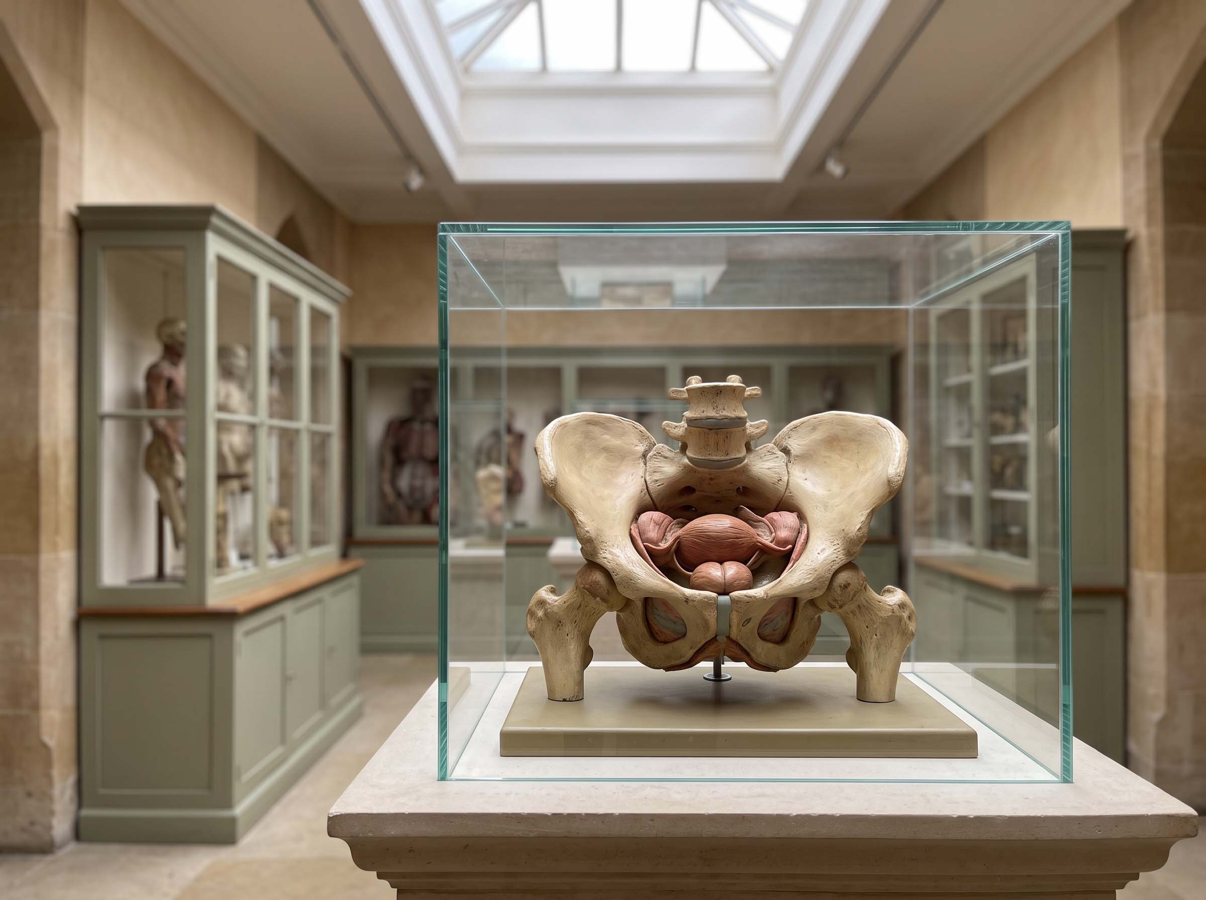
The visible portion of the clitoris is the glans, sitting beneath the prepuce at the front of the vestibule. It is a small part of a considerably larger structure. Behind the glans, the body of the clitoris runs backward before dividing into two crura (legs) that extend along the pubic arch. Flanking the vestibule on either side are the vestibular bulbs (clitoral bulbs), paired masses of erectile tissue.
In 2024, a research team at St George's University of London published the most complete pooled account of these dimensions to date. Their meta-analysis, "Beyond the tip of the iceberg: A meta-analysis of the anatomy of the clitoris" in Clinical Anatomy, followed the PRISMA protocol across the Ovid Medline and Embase databases and pooled measurements taken by MRI, ultrasound, and cadaveric dissection, as well as measurements from living women. Twenty-one studies met the inclusion criteria (Longhurst et al., 2024).
The averages they calculated make the proportions plain. The iceberg metaphor in their title is not rhetorical flourish; it is a description of the shape of the data.
| Structure | Average length | Average width |
|---|---|---|
| Glans (visible tip) | 6.40 mm (0.25 in) | 5.14 mm (0.20 in) |
| Body | 25.46 mm (1.00 in) | 9.00 mm (0.35 in) |
| Crura (paired legs) | 52.41 mm (2.06 in) | 8.71 mm (0.34 in) |
| Vestibular bulbs | 52.00 mm (2.05 in) | 10.33 mm (0.41 in) |
| Prepuce (hood) | 23.19 mm (0.91 in) | — |
Pooled averages from 21 studies using MRI, ultrasound, cadaveric dissection, and measurement in living women. Individual measurements in the underlying studies span considerably wider ranges.
Pooled average lengths in millimetres. Source: Longhurst et al., Clinical Anatomy, 2024.
The same meta-analysis documented the average distance from the clitoris to the external urethral meatus at 22.27 mm (0.88 in), to the vagina at 43.14 mm (1.70 in), and to the anus at 76.30 mm (3.00 in). Those three numbers are the reason the older habit of drawing the clitoris as a dot beside the urethra was misleading: the organ has a footprint, and the surrounding structures sit in measurable relation to it.
The glans is roughly a quarter of an inch. The crura it sits on average just over two. The visible part was never the whole organ — the drawings simply stopped there.
Why the picture took so long to assemble

Anatomical knowledge is built on donated bodies, and for most of the discipline's history the bodies dissected were overwhelmingly male. Andreas Vesalius, whose 1543 De humani corporis fabrica founded modern anatomy, treated the clitoris as an anomaly rather than a standard structure. Georg Ludwig Kobelt published detailed and largely accurate descriptions of the full clitoral complex in the 1840s, and that work then failed to carry forward into general reference texts with any consistency.
The most-cited illustration of the gap is Gray's Anatomy. The clitoris appeared in the 1901 edition and was absent from the 25th edition of 1948, reappearing in later decades in a reduced form. Analyses of contemporary teaching texts continue to find thin coverage: a University of Melbourne review of anatomy textbooks reported that most included only a single image of the vulva, often a black-and-white line drawing, that one text carried no vulval image at all, and that where inner-lip protrusion was pictured, the accompanying text usually did not mention that labial size and symmetry vary.
The practical consequence shows in the size of the evidence base. Longhurst's team, searching two major medical databases without date restriction, found 21 studies that met their criteria. For comparison, single meta-analyses of male genital dimensions have pooled samples in the tens of thousands. Anatomical detail of this kind comes largely from cadaveric dissection and imaging of volunteers, and the supply of donated female bodies is the constraint on how quickly the picture fills in. Body donation programs at teaching universities are the mechanism through which that changes; the shortfall is a supply problem before it is anything else.
Variation, and what women say about it
The measurement literature is consistent on one point: there is no single configuration that the studied populations cluster tightly around. A cross-sectional study of 200 premenopausal women attending gynecological appointments for unrelated reasons recorded a median inner-lip width of 19.0 mm (0.75 in) with an interquartile range of 12.6 to 27.5 mm, and a median length of 35.5 mm (1.40 in) with an interquartile range of 27.8 to 48.9 mm.
A separate cross-sectional study found that 56% of participants had visible labia minora and 86.5% considered their own appearance to be within the ordinary range — including two-thirds of the women in the largest measurement group. It is honest to record both halves of that finding. Most women in the sample were untroubled by their appearance, and a minority were not; among those who described their genitals as unusual, 73.3% had visible inner lips.
Preferences exist, they are documented, and pretending otherwise does not serve readers. What the anatomical evidence does establish is that the visible range in the published measurement studies is much wider than the range depicted in either textbooks or commercial imagery, and that researchers describing these cohorts use the language of variation rather than the language of deviation.
Did You Know?
In Kreklau's 2018 cohort of 657 women aged 15 to 84, inner-lip length showed a statistically significant inverse correlation with age (r = −0.364, p < 0.001) — one of the few lifespan patterns in vulval anatomy documented at that sample size. The study's authors noted their sample was deliberately drawn from a single ethnic group, which limits how far the figures generalize.
The G-spot: what the anatomical literature actually reports
Few topics in this field carry more popular certainty and less anatomical agreement. The published debate is narrow and specific, and worth summarizing on its own terms.
In 2012, Adam Ostrzenski reported finding a discrete structure during the dissection of a single cadaver, describing it as having three sections with a fibroconnective capsule and apparently erectile contents, measuring around 8 mm at its longest dimension and typically sitting off the midline. In 2017, a team including the urologist Helen O'Connell published systematic cadaveric dissections of the anterior vaginal wall in The Journal of Sexual Medicine and reported no macroscopic structure at the proposed location. What they did describe was a merging of the anterior aspect of the distal vaginal wall with the clitoris, to the point that separating clitoris, urethra, and vagina at that junction was difficult (Hoag, Keast, and O'Connell, 2017).
A 2021 systematic review in Sexual Medicine concluded that the studies do not agree on whether the structure exists, and that among those treating it as real there was no agreement on its location, size, or nature. Several authors have proposed that the region is better described as the clitourethrovaginal complex — a functional relationship between the internal clitoris, the urethra, and the anterior vaginal wall rather than a discrete organ. That framing is consistent with the clitoral dimensions in Table 1: the crura and bulbs sit exactly where a separate structure was being sought.
Estrogen and the tissue across a lifespan
Vulval and vaginal tissue is hormone-responsive, and its structure changes with circulating estrogen rather than staying fixed from puberty onward. Before puberty, these tissues are thin and unstimulated. Rising estrogen at puberty thickens the vaginal epithelium, increases glycogen content in its cells, promotes fat deposition in the mons pubis and labia majora, and enlarges the labia minora and clitoral structures. Cyclical hormonal change continues through the reproductive years — one of the studies included in the Longhurst meta-analysis specifically examined morphometric and vascular changes in the clitoris across the menstrual cycle (Battaglia et al., The Journal of Sexual Medicine, 2008). After menopause, falling estrogen reverses much of the pubertal pattern. On the specific question of clitoral glans width after menopause, the studies pooled in the 2024 meta-analysis did not find a significant difference, though one included study did record a difference in the distance between the clitoris and the urethral meatus. For the wider picture of how hormones shape body composition and development, see our reference on hormones and body development.
Shared origins with male anatomy
Every structure named in this guide has a counterpart in male anatomy, because both develop from the same embryonic tissue before hormonal signals differentiate them. The genital tubercle becomes either the glans of the clitoris or the glans of the penis. The urogenital folds become the labia minora or fuse to form the penile shaft. The labioscrotal swellings become the labia majora or fuse into the scrotum. The vestibular bulbs correspond to the bulb of the penis, and the crura to the crura of the penis.
This is why the two sets of structures share erectile tissue, nerve supply patterns, and general architecture, and why the equivalent structures in male anatomy are described using much of the same vocabulary — a comparison set out in detail in our guide to how the penis works. It is also the strongest argument against treating female external anatomy as a footnote to the male version. The developmental logic runs in parallel, not in sequence.
| Embryonic structure | Develops into (female) | Develops into (male) |
|---|---|---|
| Genital tubercle | Glans of the clitoris | Glans of the penis |
| Urogenital folds | Labia minora | Ventral penile shaft (fused) |
| Labioscrotal swellings | Labia majora | Scrotum (fused) |
| Urogenital sinus | Vestibule, lower vagina, urethra | Prostatic and membranous urethra |
| Erectile primordia | Crura and vestibular bulbs | Crura and bulb of the penis |
The vaginal canal and cervix, briefly
Past the introitus, the vagina is a muscular canal lined with the same estrogen-responsive epithelium described above. At its upper end sits the cervix (neck of the uterus), projecting into the canal and surrounded by recesses called the fornices. One measurement study of 208 women recorded average anterior fornix depth at 7.75 cm (3.05 in) and posterior fornix depth at 9.25 cm (3.64 in) — a reminder that the canal is not symmetrical front to back. Beyond that boundary, the anatomy belongs to a different reference; the point here is only that the vulva ends and the vagina begins at a specific, nameable place.
Naming things accurately is the whole exercise
An educated reader should be able to name a dozen structures on the outside of the female body and say roughly where each one sits, what tissue it is made of, and how much it varies between individuals. That is an ordinary expectation for any other region of anatomy. The reason it has not been an ordinary expectation here is a documented history of thin illustration, inconsistent labeling, and a small evidence base — all of which are correctable, and some of which has already been corrected. Terms used here are defined in the Anatomy Glossary.
Questions Readers Ask
Sources
- Longhurst, G.J., Beni, R., Jeong, S.R., et al. "Beyond the tip of the iceberg: A meta-analysis of the anatomy of the clitoris." Clinical Anatomy, 37(2), 233–252, 2024.
- Kreklau, A., Vâz, I., Oehme, F., et al. "Measurements of a 'normal vulva' in women aged 15–84: a cross-sectional prospective single-centre study." BJOG, 2018.
- Lloyd, J., Crouch, N.S., Minto, C.L., Liao, L.M., Creighton, S.M. "Female genital appearance: 'Normality' unfolds." BJOG, 112(5), 643–646, 2005.
- Hoag, N., Keast, J.R., O'Connell, H.E. "The 'G-Spot' Is Not a Structure Evident on Macroscopic Anatomic Dissection of the Vaginal Wall." The Journal of Sexual Medicine, 14(12), 2017.
- Vieira-Baptista, P., et al. "G-spot: Fact or Fiction? A Systematic Review." Sexual Medicine, 9(5), 2021.
- Ostrzenski, A. "G-spot anatomy: A new discovery." The Journal of Sexual Medicine, 9, 1355–1359, 2012.
- Battaglia, C., et al. "Menstrual cycle-related morphometric and vascular modifications of the clitoris." The Journal of Sexual Medicine, 5, 2853–2861, 2008.
- Systematic Review and Meta-Analysis of Labia Minora Anatomy in Premenopausal Women, 2026 (seven studies, 991 women).
- University of Melbourne review of vulval representation in anatomy textbooks.
Disclaimer
- All content on this website—including articles, educational materials, and interactive calculators—is for informational, educational, and entertainment purposes only. The calculations, percentiles, and outputs generated by tools on this site are based on general statistical data and mathematical models; they do not constitute medical data, a clinical assessment, or a diagnosis.
- Nothing contained on this website is a substitute for professional medical advice, diagnosis, or treatment. Always seek the guidance of a qualified healthcare professional or urologist with any questions you have regarding physical development, anatomy, or health conditions. Reliance on any information or tools provided by this website is solely at your own risk.


