



 English
 Español


At six weeks in, the blueprint is identical after fertilization, a human embryo is roughly 10 to 14 mm long (about 0.4 to 0.6 in) — the length of a coffee bean. Along the back wall of its abdominal cavity sit two small ridges of tissue.
Below, on the outside, there is a rounded bud, two folds of tissue flanking a shallow groove, and two swellings on either side of that. Every human body has had these structures. Nothing about them, at that moment, indicates what they will become. The external and internal female structures catalogued in adult anatomy references and the male structures beside them in the same textbook are, at six weeks, the same three or four pieces of tissue.
Anatomists have a name for this period: the indifferent stage. The ridges are called bipotential gonads, meaning they carry the machinery to become either testes or ovaries and have not yet committed. The bud on the outside is the genital tubercle. The folds and swellings around it are the urogenital folds and the labioscrotal swellings. Alongside the gonads run two complete sets of plumbing — the Wolffian ducts and the Müllerian ducts — and at this stage every embryo has both.
What happens next is one of the most precisely ordered sequences in human biology, and it is worth understanding on its own terms rather than as a story about which body came first.
In Brief
- Every human embryo passes through a stage in which the reproductive structures are identical and undifferentiated.
- A single gene begins the divergence, but ovary formation is now understood to be an actively driven program of its own, not an absence of instructions.
- Three hormonal signals — anti-Müllerian hormone, testosterone and dihydrotestosterone — each have separate, specific jobs.
- Research points to a narrow first-trimester window in which timing matters more than quantity.
- Published pediatric research describes puberty as a sequence with wide age ranges, and anatomy continues to change well past it.
The difference between male and female anatomy is not a difference of parts. It is a difference of instructions given to the same parts.
One switch — and a second program running alongside it
The divergence begins with a gene on the Y chromosome. Identified in 1990 by two research groups working on the same problem, it was named SRY, for sex-determining region of the Y chromosome. Its expression in the bipotential gonad is brief and early, and it does one thing: it starts a cascade that turns the supporting cells of the ridge into Sertoli cells, committing the tissue to becoming a testis. A review in Physiological Reviews describes this decision as the hinge on which the rest of the sequence turns — once the gonad is committed, the hormones it makes govern everything downstream.
The older textbook account stopped there and called female development the "default" pathway. That framing came from a genuinely elegant experiment. In 1947 the French embryologist Alfred Jost removed the gonads from rabbit fetuses before they had differentiated, then returned them to the uterus. Every one of them — regardless of chromosomes — developed the female pattern of ducts and external structures. Jost concluded that the testis actively imposes one outcome and that the other happens on its own.
Research since the mid-2000s has revised the second half of that conclusion. Studies of the genes WNT4, RSPO1 and FOXL2 show that ovary formation is driven by its own coordinated program, one that has to actively suppress the testis pathway rather than simply lacking it. The most striking demonstration came in 2009, when researchers reporting in Cell switched off FOXL2 in the ovaries of adult mice and watched ovarian supporting cells reorganize into cells resembling those of a testis. The implication is that in females the ovarian program is not a decision made once in the first trimester. It is a signal that keeps being sent for life.
Did You Know?
For roughly sixty years, the phrase "female development is the default" appeared in nearly every embryology textbook. It survives in popular science writing today. The laboratory finding that overturned it was published in 2009, and the correction has not fully reached general audiences — a reminder that developmental biology is an active research field, not a closed subject.
Three signals, three separate jobs
Once a testis forms, it produces two hormones with entirely different targets, and a third molecule is made elsewhere from one of them. Reading these as a single force called "male hormones" hides how specific each one is.
Anti-Müllerian hormone is released by Sertoli cells from around the seventh to eighth week. Its only job in this sequence is subtraction: it causes the Müllerian ducts to regress. Without it, those ducts remain and go on to form the uterine tubes, the uterus and the upper portion of the vagina.
Testosterone comes from Leydig cells, which appear at around the ninth week. It stabilizes the Wolffian ducts, which would otherwise disappear, and directs them toward becoming the epididymis, the vas deferens and the seminal vesicles. Jost's later work separated these two effects clearly — testosterone builds one duct system, and a different testicular factor removes the other.
Dihydrotestosterone, usually shortened to DHT, is not made in the testis. It is converted from testosterone by an enzyme called 5-alpha-reductase in the tissues of the external structures themselves. DHT is what drives the genital tubercle to lengthen, the urogenital folds to fuse along the midline into the shaft and its urethral channel, and the labioscrotal swellings to fuse into the scrotum. It also directs formation of the prostate. Anyone curious about how the finished structures work can follow that thread into how the adult male anatomy is organized.
In the female sequence, estrogen produced by the fetal and maternal environment supports the development of the Müllerian-derived structures, while the same tubercle, folds and swellings develop without fusion: the tubercle becomes the clitoris, the urogenital folds remain separate as the labia minora, and the labioscrotal swellings remain separate as the labia majora. The word "remain" is doing real work there — these are not incomplete versions of anything. They are the unfused configuration of the identical starting tissue.
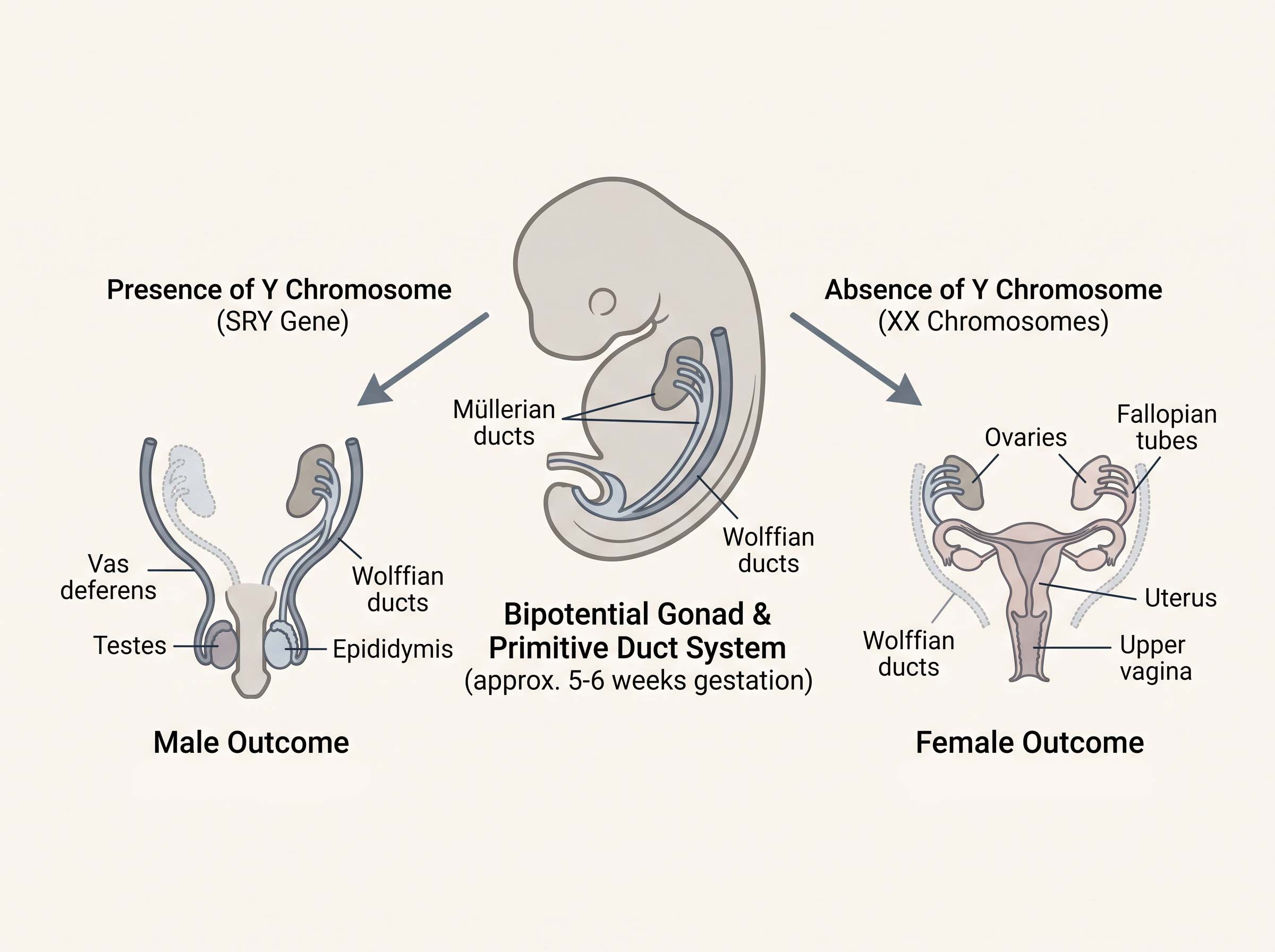
The same tissue, two destinations
Because the starting material is shared, adult structures line up in pairs. Anatomists call these homologues — structures with a common embryonic origin, whatever their eventual size, position or function. The table below sets out the main correspondences.
| Embryonic structure | Develops into (male pattern) | Develops into (female pattern) |
|---|---|---|
| Bipotential gonad | Testis | Ovary |
| Genital tubercle | Glans and shaft of the penis | Clitoris (glans and body) |
| Urogenital folds | Fuse to enclose the urethral channel | Labia minora (remain unfused) |
| Labioscrotal swellings | Scrotum | Labia majora |
| Wolffian ducts | Epididymis, vas deferens, seminal vesicles | Regress |
| Müllerian ducts | Regress | Uterine tubes, uterus, upper vagina |
| Gubernaculum | Guides descent of the testes | Ovarian and round ligaments |
Correspondences as set out in standard embryology references; the male and female columns describe two outcomes of one shared starting structure, not a ranking.
Timing is the hidden variable
One of the least-discussed findings in this field concerns when hormonal signals arrive rather than how much of them there is. Working with rats, Welsh and colleagues reported in 2008 in the Journal of Clinical Investigation that androgen action had to occur within a specific early window in order for the reproductive tract to develop along the male pattern. Blocking that action inside the window produced lasting differences; altering hormone levels afterward did not. They named it the masculinization programming window and estimated, from the timings observed, that the equivalent period in humans is likely between 8 and 14 weeks of gestation.
What makes this striking is that the window closes before the external structures visibly differentiate at all. The instructions are issued first; the anatomy follows later. Researchers have proposed anogenital distance — the measured span between two fixed landmarks — as a readout of hormone exposure during that window, since in human studies the male–female difference in that measurement first emerges at around 11 to 13 weeks and widens through the second trimester. The evidence base in humans is indirect and drawn largely from animal models, and researchers writing on the subject describe further work as needed. It remains among the clearer illustrations of a general principle: some developmental processes are time-sensitive in ways that are permanent.
The puberty nobody remembers
Sexual development is not one event with a long pause before it. In the first months after birth, the hormone axis that controls reproduction switches on briefly, then goes quiet again. Kuiri-Hänninen, Sankilampi and Dunkel, reviewing this phase in 2014, describe testosterone in infant boys rising to concentrations that peak at roughly one to three months of age before subsiding, and hormone activity in infant girls persisting in a fluctuating pattern for considerably longer. Researchers call it mini-puberty. Its full purpose is still being worked out, and it is one of the reasons developmental biologists describe this system as running on a schedule rather than sitting dormant. Readers following the endocrine side of this can go deeper in the site's hormones and body development section.
Puberty is a sequence, not a schedule
The reference framework still used to describe pubertal change comes from a longitudinal study conducted in England by W. A. Marshall and J. M. Tanner, published in two papers in 1969 and 1970. Their contribution was staging — a way of describing where a body is in the sequence rather than assigning a single expected age.
What the published data show is that the order is fairly consistent and the timing is not. In girls, the typical first visible change is breast budding, followed roughly one to one and a half years later by pubic hair development, with first menstruation arriving on average some two to two and a half years after breast budding. In boys, the first change is enlargement of the testes, followed by growth of the penis and then pubic hair; the growth spurt arrives comparatively later in the sequence than it does in girls. Standard pediatric summaries place the onset range at roughly 8 to 13 years in girls and 9 to 14 years in boys — a five-year span on either side, all of it within the studied range.
Those ranges are not fixed across generations either. A 2020 systematic review and meta-analysis published in JAMA Pediatrics pooled thirty studies and found that the average age at breast development declined by about 0.24 years — nearly three months — per decade between 1977 and 2013. The authors noted that this shift may make older diagnostic age thresholds outdated in some regions. Proposed explanations include nutrition, body composition and environmental exposures, and the relative contribution of each remains an open research question.
Figure 1 — Approximate age ranges reported in published pediatric research for the appearance of each pubertal milestone. Bars show ranges, not averages; individual sequences vary in both timing and pace.
The blueprint keeps working
Adult anatomy is often written about as a finished state. The research does not describe it that way. In women, the fall in estrogen around and after menopause is associated with documented changes in the tissues of the vulva and vagina — reviews in the endocrinology literature describe thinning of the surface layer, reduced elasticity as elastin and collagen are reorganized, and reduced blood supply. Researchers writing on the subject are candid that the mechanisms connecting hormone levels to specific tissue outcomes are still incompletely mapped.
In men, hormone change is gradual rather than staged. Harman and colleagues, reporting on the Baltimore Longitudinal Study of Aging in 2001, found declines in total and free testosterone with advancing age when the same men were followed over time; Feldman and colleagues, publishing longitudinal results from the Massachusetts Male Aging Study in 2002, reported a fall in total testosterone of roughly 0.8 percent per year in middle age, with a steeper decline in the free fraction. These are population-level averages drawn from research cohorts, and individual figures vary widely.
The pattern worth taking from both is the same one that governs the fetal sequence: this tissue responds to its hormonal environment throughout life, not only at the start of it.
When the sequence runs differently
Because each step in the cascade is specific, an alteration at any one point produces a specific outcome rather than a general one. Conditions grouped under the term differences of sexual development have been studied for exactly this reason — they have shown researchers which signal does which job. The best-known example concerns 5-alpha-reductase, the enzyme that converts testosterone into DHT. Studies of communities where a variant affecting that enzyme is more common, first documented by Imperato-McGinley and colleagues in 1974, established that DHT rather than testosterone itself directs the development of the external structures. That single finding sits behind a large part of what is now standard in embryology texts.
These are developmental variations, not the subject of this article, and they are described here only because they explain how the mechanism was worked out. This site does not offer diagnostic or clinical guidance of any kind.
What the shared origin actually tells us
The useful thing about starting an anatomy education at the indifferent stage is that it reorders everything that follows. Male and female structures stop looking like two separate systems that happen to sit in the same region of the body, and start looking like what they are — one set of tissues that received different instructions at different moments, on a schedule precise enough that a two-week shift in timing leaves a permanent mark, and loose enough that the age at which any given person's puberty begins can vary by five years without leaving the studied range.
The bud, the folds, the swellings and the ridge. Every human body has carried them. What each one became is a matter of which signals arrived, and when.
Editorial Glossary
- Bipotential gonad
- The early ridge of tissue that can still become either a testis or an ovary.
- Genital tubercle
- The small external bud present in every embryo that later becomes either the penis or the clitoris.
- Homologue
- A structure that shares a common embryonic origin with another, whatever its final form.
- Indifferent stage
- The period during which reproductive structures are present but not yet distinguishable.
- Sensitive window
- A limited period during which a signal must arrive to have its effect; the same signal later produces a different result or none.
- Staging
- Describing where a body is in a developmental sequence rather than assigning it an expected age.
Common Questions
Editorial note: This article describes typical developmental sequences and the age ranges reported in published research. It is educational reference material. It is not diagnostic, does not describe any individual case, and offers no clinical or personal guidance.
Sources and further reading
- Jost, A. (1947). Recherches sur la différenciation sexuelle de l'embryon de lapin. Archives d'Anatomie Microscopique et de Morphologie Expérimentale, 36, 271–315.
- Sinclair, A. H. et al. (1990). A gene from the human sex-determining region encodes a protein with homology to a conserved DNA-binding motif. Nature, 346, 240–244.
- Wilhelm, D., Palmer, S. & Koopman, P. (2007). Sex determination and gonadal development in mammals. Physiological Reviews, 87(1), 1–28.
- Uhlenhaut, N. H. et al. (2009). Somatic sex reprogramming of adult ovaries to testes by FOXL2 ablation. Cell, 139(6), 1130–1142.
- Welsh, M. et al. (2008). Identification in rats of a programming window for reproductive tract masculinization. Journal of Clinical Investigation, 118(4), 1479–1490.
- Sharpe, R. M. (2020). Androgens and the masculinization programming window: human–rodent differences. Biochemical Society Transactions, 48(4), 1725–1735.
- Kuiri-Hänninen, T., Sankilampi, U. & Dunkel, L. (2014). Activation of the hypothalamic-pituitary-gonadal axis in infancy: minipuberty. Hormone Research in Paediatrics, 82(2), 73–80.
- Marshall, W. A. & Tanner, J. M. (1969, 1970). Variations in the pattern of pubertal changes in girls; and in boys. Archives of Disease in Childhood, 44 & 45.
- Eckert-Lind, C. et al. (2020). Worldwide secular trends in age at pubertal onset assessed by breast development among girls. JAMA Pediatrics, 174(4), e195881.
- Harman, S. M. et al. (2001). Longitudinal effects of aging on serum total and free testosterone levels in healthy men. Journal of Clinical Endocrinology & Metabolism, 86(2), 724–731.
- Feldman, H. A. et al. (2002). Age trends in the level of serum testosterone and other hormones in middle-aged men. Journal of Clinical Endocrinology & Metabolism, 87(2), 589–598.
- Nappi, R. E. et al. (2019). Addressing vulvovaginal atrophy for healthy aging in women. Frontiers in Endocrinology, 10, 561.
- Imperato-McGinley, J. et al. (1974). Steroid 5-alpha-reductase deficiency in man. Science, 186(4170), 1213–1215.
- Laqueur, T. (1990). Making Sex: Body and Gender from the Greeks to Freud. Harvard University Press.
Disclaimer
- All content on this website—including articles, educational materials, and interactive calculators—is for informational, educational, and entertainment purposes only. The calculations, percentiles, and outputs generated by tools on this site are based on general statistical data and mathematical models; they do not constitute medical data, a clinical assessment, or a diagnosis.
- Nothing contained on this website is a substitute for professional medical advice, diagnosis, or treatment. Always seek the guidance of a qualified healthcare professional or urologist with any questions you have regarding physical development, anatomy, or health conditions. Reliance on any information or tools provided by this website is solely at your own risk.


